Mayo Clinic’s AI for Early Cancer Detection Turns Routine CT and MRI into an Early‑Warning System
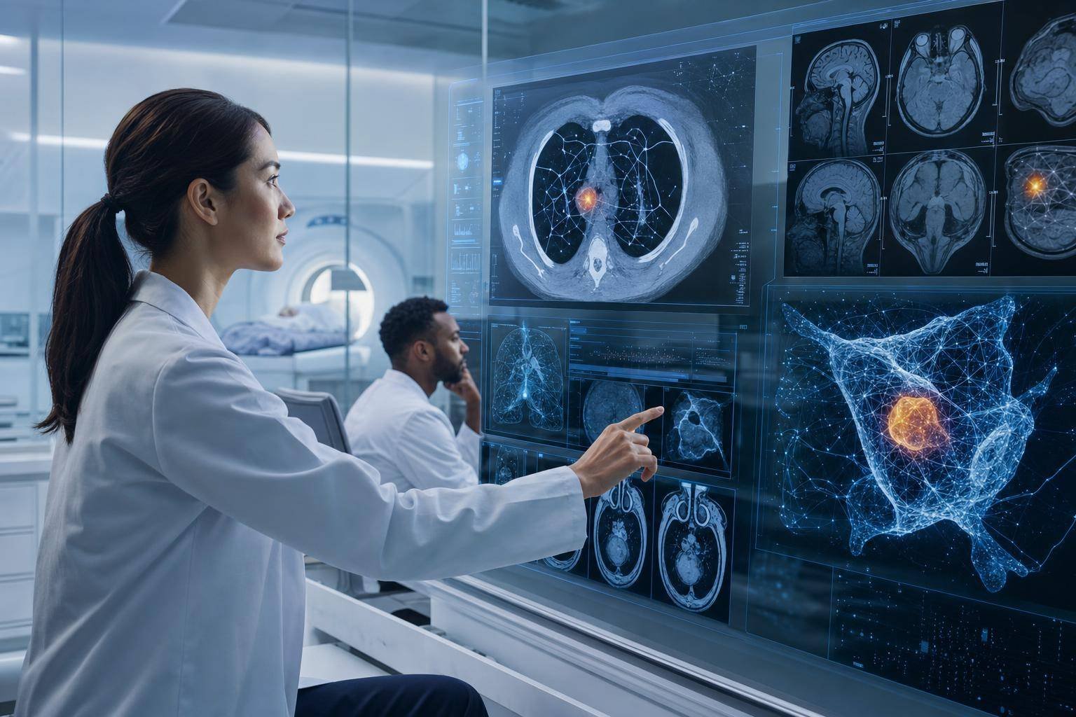
Medical imaging already sees what the naked eye can’t. Now, Mayo Clinic researchers say it can sense what even trained radiologists often won’t spot until months later. Their newly announced AI system analyzes routine CT and MRI scans for faint, early cancer signatures—surfacing subtle patterns long before disease becomes clinically obvious.
Why now? Screening and diagnosis are under acute pressure: volumes are up, radiology teams are stretched, and many cancers still present late. An AI that quietly triages studies in the background, flags suspicious regions, and helps standardize quality could be the rare win that improves patient outcomes and relieves clinical load without disrupting existing workflows.
Below, we break down what Mayo built, how it works technically, why it matters clinically, and what health systems should do to responsibly adopt AI for early cancer detection—without drifting into hype or overlooking hard regulatory, security, and bias challenges. For additional context, see Mayo’s announcement and research summary: Mayo Clinic researchers develop AI model for early cancer detection.
What Mayo Clinic Built: An AI Early Cancer Detection Model for Routine Imaging
Mayo Clinic’s team trained a deep learning model to identify subtle lesion patterns on CT and MRI—especially in the lung, liver, and pancreas. Those organs are high-stakes: earlier detection can dramatically change prognosis, yet current screening coverage and sensitivity can be limited. The model was trained on millions of de-identified scans from across Mayo’s global network, then tested retrospectively. In a significant share of cases, the system flagged malignancies months before they would have otherwise been detected, while keeping false positives low enough to be clinically useful.
Critically, this is not a “diagnose-and-discharge” bot. It’s a triage co-pilot that:
- Runs in the background inside hospital infrastructure
- Ranks studies that may warrant urgent review
- Produces heatmaps to guide a radiologist’s attention
- Leaves final interpretation fully in human hands
Early pilot feedback suggests the assistance can reduce reader fatigue and help standardize quality across high-volume settings. That “assistive, not autonomous” posture matches how successful radiology AI is being integrated elsewhere—supporting real-world needs without ripping and replacing established clinical processes.
Target cancers and modalities
- Lung: The system evaluates chest CTs, including low-dose scans commonly used for screening. Early pulmonary nodules can be hard to assess at first glance, especially across dense daily worklists.
- Liver and pancreas: Abdominal CT and MRI provide rich 3D information, but small or isoattenuating lesions can be nuanced. An extra set of (machine) eyes is appealing in hepatobiliary and pancreatic disease, where early intervention matters.
Training data and validation
The team trained on large, diverse archives of de-identified imaging studies. De-identification was done in alignment with HIPAA’s safe harbor and expert determination pathways (HHS HIPAA de-identification guidance). Retrospective validation demonstrated earlier flagging of malignancies in a meaningful fraction of cases, with false-positive rates low enough to avoid overwhelming clinicians. Prospective trials are planned to verify generalizability, real-world impact, and safety.
Designed to augment radiologists
AI outputs include:
- Case-level triage scores to prioritize reading order
- Region-level heatmaps that highlight suspicious tissue
- Uncertainty measures that can indicate when caution is warranted
- Structured metadata to support auditability and quality improvement
Radiologists retain control of thresholds, can dismiss or accept AI prompts, and can use AI overlays as decision support rather than directives. That human-in-the-loop design is essential to mitigate automation bias and maintain clinical standards.
Under the Hood: How Deep Learning Finds Subtle Cancer Signals
While Mayo has not published every architectural detail, state-of-the-art imaging AI typically relies on a toolkit of techniques that fits this use case:
- 3D convolutional neural networks (3D CNNs): Capture spatial context across slices, essential for differentiating real lesions from noise or vessel cross-sections.
- Vision transformers (ViTs): Self-attention layers can model long-range dependencies and heterogeneous textures across the volume.
- Multi-instance learning: Useful when labels are at the study or organ level rather than pixel-perfect annotations; the model learns which patches signal disease.
- Preprocessing and harmonization: Intensity normalization, resampling to standardized voxel sizes, and metal artifact mitigation help reduce scanner and protocol variability.
- Contrast-phase awareness: For multi-phase CT/MRI, the model may incorporate contrast timing to better distinguish vascular structures from lesions.
- Saliency and explainability: Gradient-based methods (e.g., Grad-CAM) and occlusion tests highlight model-sensitive regions, aligning AI guidance with radiologists’ interpretive workflow.
- Uncertainty estimation: Techniques like test-time augmentation, Monte Carlo dropout, or shallow ensembles yield confidence measures that can inform triage thresholds.
The central challenge is sensitivity without alert fatigue. If an AI pings too many benign anomalies, clinicians ignore it. If it’s too conservative, it misses the point of early detection. Practical deployment therefore leans heavily on calibration: adjusting decision thresholds to local prevalence, scanner mix, and reader preferences. Simple but robust methods like temperature scaling can improve probability accuracy so that “90% confidence” behaves like 90% in the real world.
Clinical Performance, Validation, and the Hype-to-Reality Gap
Early-detection AI is tantalizing because downstaging disease saves lives and costs. But it also lives at the edge of the signal-to-noise envelope. This is where rigorous validation matters.
- Retrospective vs. prospective: Retrospective studies can overstate performance if training and test distributions overlap (same scanners, sites, protocols). Prospective trials, ideally randomized or with stepped-wedge designs, determine if earlier flags actually change outcomes.
- Population and scanner diversity: Performance can drift across demographics, body habitus, comorbidities, and vendor-specific imaging pipelines. External validation across multiple centers is a must.
- Metrics that matter: Beyond AUC and sensitivity/specificity, measure positive predictive value (PPV), false-positive rate per study, time-to-diagnosis gains, and downstream utilization (e.g., unnecessary follow-up imaging).
- Reader–AI synergy: Evaluate double-reading models, how juniors vs. seniors benefit, and whether AI helps standardize across sites. Radiology societies such as the RSNA track emerging evidence and practical guidance for AI-Augmented reading (RSNA AI resources).
Context is key: screening norms and evidence vary by cancer type. For lung cancer, for example, recommended screening with low-dose CT has structured protocols and lexicons (e.g., Lung-RADS) to reduce variability. Any AI assist should align with these frameworks, not invent new categories out of thin air (ACR lung cancer screening resources).
Finally, the benefit must outweigh the burden. If AI prompts increase follow-up scans and biopsies without improving outcomes, it’s a net negative. The goal isn’t merely to find more “things” but to find the right things earlier—things that, when acted upon, lead to better survival and quality-adjusted life years. The National Cancer Institute maintains accessible overviews of screening rationales and uncertainties that can anchor institutional conversations about where AI helps most (NCI overview of cancer screening).
Privacy, Security, and Regulatory Guardrails
Mayo’s announcement makes the right noises—and from what’s described, meaningful commitments—on governance:
- Data governance and de-identification: Training data were de-identified per HIPAA, using controlled processes and audit logging. This reduces privacy risk but does not eliminate it; research teams must continue to watch for re-identification pathways (e.g., rare pathologies or device signatures).
- Network architecture: The model runs within secure hospital infrastructure, with strict role-based access controls, logging, and no direct connection to external model providers. This limits third-party exposure and supports incident response.
- Change control and monitoring: Versioning models, documenting training lineages, and capturing inference metadata (software version, thresholds, DICOM attributes) are crucial to traceability.
Two external frameworks can help organizations translate principles into practice:
- NIST AI Risk Management Framework (AI RMF): A comprehensive, vendor-neutral guide for mapping, measuring, and managing AI risks across the lifecycle—from data to deployment to monitoring. It’s increasingly used by healthcare CISOs, compliance leaders, and AI governance boards (NIST AI RMF 1.0).
- FDA pathway for AI/ML-enabled Software as a Medical Device (SaMD): Clinical AI that influences diagnosis or treatment typically requires regulatory clearance. While triage aids may fall into different classes depending on claims, any large-scale clinical deployment should expect to align with FDA expectations for safety, effectiveness, labeling, and post-market surveillance (FDA AI/ML SaMD overview).
Health systems should assume regulators will also scrutinize cybersecurity controls (e.g., SBOMs, patch management, third-party risk) and post-market real-world performance reporting. AI that touches patient care must be treated with the same rigor as other clinical devices.
Implementing AI Triage in Radiology: A Practical Playbook
Hospitals don’t need to reinvent the wheel to pilot AI for early cancer detection. They do need a thoughtful plan that blends clinical leadership, IT, security, and data science.
1) Define the clinical objective and guardrails – Decide where AI can add clear value: e.g., prioritizing chest CTs with suspicious nodules, surfacing small hepatic lesions on abdominal MRI. – Pre-commit to success metrics: reduction in time-to-review for positive cases, reader agreement, PPV at chosen thresholds, and net impact on downstream utilization. – Establish initial exclusion criteria (e.g., pediatric studies, certain contrast protocols) if model performance is unproven in those populations.
2) Stand up governance and risk management – Form a cross-functional committee: radiology leadership, oncology, IT/PACS, security, compliance, and frontline clinicians. – Adopt an AI risk framework (e.g., NIST AI RMF) and document accountability: who approves thresholds, who monitors drift, who responds to incidents. – Require model cards and datasheets: training sources, known limitations, intended use, performance by subgroup, and failure modes.
3) Prepare technical integration the right way – PACS and DICOM: Ensure bidirectional integration that can fetch studies, write back overlays, and preserve original images. Work with your vendor to validate DICOM conformance (DICOM Standard). – Workflow events via HL7/FHIR: Trigger AI inference when new studies arrive, and route results to the right worklists. Use interoperable schemas to avoid brittle one-offs (HL7 FHIR overview). – Compute and latency: AI should finish inference before a typical read starts. Profiling and, if needed, GPU acceleration on-prem compute can help. – Fail-safe behavior: If AI is down, radiology workflows must continue uninterrupted.
4) Design a pilot that learns fast – Pick a limited set of scanners and protocols with clear ground truth pathways. – Calibrate thresholds locally: Start with conservative triage, then adjust to balance sensitivity and alert volume. – Shadow mode first: Run AI without influencing reads to establish baseline performance. Then move to assisted reads with clear instructions.
5) Train and support your clinicians – Short, focused training on how to interpret heatmaps, handle uncertainty flags, and avoid automation bias. – Encourage feedback loops: A one-click “useful/not useful” prompt and error tagging helps drive iterative improvement.
6) Monitor relentlessly – Daily and weekly dashboards: case counts, alert rates, PPV/NPV estimates from chart review samples, turnaround times, and overrides. – Drift detection: Watch for distribution shifts in scanner metadata, image statistics, and outcome prevalence. Recalibrate or retrain promptly. – Safety signals: Track unanticipated adverse events (e.g., inappropriate follow-ups) and near-misses.
Mistakes to avoid
- Deploying without a clear intended use and success metrics
- Letting vendors set thresholds without local calibration
- Ignoring user experience—buried overlays and confusing scores are functionally useless
- Underinvesting in validation across your scanner fleet and protocols
- Failing to plan for version upgrades, rollbacks, and audit trails
- Treating explainability as a checkbox rather than a training and trust-building tool
How the Model Augments Human Judgment—With Real-World Examples
- Pancreas “before it blooms”: Small hypoenhancing pancreatic lesions can hide in plain sight on early-phase imaging. AI heatmaps that consistently flag the same tail-of-pancreas area across timepoints prompt a second, more careful read and, if warranted, earlier MRCP or EUS referral.
- Liver lesions in complex livers: In patients with steatosis or cirrhosis, texture cues can be misleading. A calibrated AI may surface atypical patterns that merit a LI-RADS category recheck—without claiming diagnostic finality.
- Chest CT triage during peak hours: When 50 studies hit the queue at once, any tool that pushes the 3–5 most suspicious to the top can shave hours off time-to-review for likely-positive patients. Even a small acceleration can matter clinically in fast-progressing cases.
These examples reflect how triage AI slots into existing lexicons and workflows. Radiologists stay in charge of categorization, reporting, and communication.
Bias, Generalization, and Performance Drift: What to Watch
Three recurring risks demand ongoing management:
- Scanner and protocol heterogeneity: Variations in kernel, slice thickness, contrast timing, or noise profiles can degrade model performance. Preprocessing pipelines help, but they’re not panaceas. Validate across your fleet and revisit when hardware or protocols change.
- Demographic and clinical diversity: Performance may differ by age, sex, race/ethnicity, comorbidities, or body habitus. Even if ground truth is scarce, sample audits can detect early warning signs of subgroup disparities.
- Temporal drift: Prevalence, referral patterns, and imaging techniques evolve. Models trained on last year’s data can get stale. Plan for periodic recalibration and, where appropriate, controlled retraining.
Mitigations include continuous QA sampling, threshold adjustments, uncertainty-aware routing (e.g., sending low-confidence cases to senior readers), and periodic external validation.
Security Considerations for Clinical AI
Even on-premises AI introduces a new software supply chain and attack surface:
- Least-privilege access: Lock down model servers, limit PHI exposure, and audit all access. Integrate with enterprise IAM and SIEM.
- Dependency hygiene: Maintain an SBOM for AI components; patch CVEs promptly; containerize for predictable deployments.
- Data integrity: Protect DICOM stores and inference pipelines against tampering. Even slight image perturbations can cause model misbehavior.
- Incident response: Predefine “AI-off” procedures so care continues smoothly if you need to suspend inference.
Security is not just about preventing breaches; it’s about ensuring dependable, predictable behavior of systems that influence medical decisions.
Beyond Detection: Multimodal AI That Fuses Imaging, EHR, Labs, and Genomics
Mayo’s team signaled ambitions to integrate imaging-derived signals with laboratory values and genomic data. Multimodal models could, for instance:
- Combine CT texture cues with CA 19-9 or AFP trends
- Factor in smoking history, family history, or prior imaging trajectories
- Use polygenic risk scores or tumor mutation signatures where clinically indicated
Potential benefits include better risk stratification, fewer false positives, and more personalized follow-up recommendations. But the risks scale, too: data governance complexity, consent and secondary-use questions, and compounding bias if one modality reflects historical inequities. Pilot conservatively, ensure transparent feature attribution where possible, and align outputs with evidence-based pathways.
Future of AI-Assisted Screening: Trends to Watch
- Self-supervised and foundation models: Pretraining on massive unlabeled imaging corpora can reduce labeled-data requirements and boost generalization.
- Federated and privacy-preserving learning: Train across sites without pooling PHI, or use synthetic data to augment rare cases—while recognizing current limits.
- Uncertainty-aware triage: Models that know when they don’t know can route edge cases to more experienced readers.
- Real-time, edge inference: GPU-accelerated appliances in the data center or even at the modality level will keep latency low and network requirements modest.
- Payment and policy: Reimbursement models for AI-assisted screening and quality improvement will influence adoption curves more than benchmarks alone.
- Standards-first integration: The health IT community will increasingly reward AI that cleanly integrates via DICOM, HL7, and FHIR—reducing total cost of ownership and vendor lock-in.
How to Evaluate a Vendor Model Today
If you’re not building in-house, borrow this short checklist for vendor evaluations:
- Intended use and claims: Are they clinically specific and aligned with your needs?
- Evidence: Do they provide external validation and, ideally, prospective or real-world performance data?
- Subgroup reporting: Performance across scanner types, protocols, and demographics
- Workflow fit: DICOM overlays, worklist integration, and reader-friendly UX
- Governance artifacts: Model card, change-control policy, audit logs, and rollback plan
- Security posture: SBOMs, pentest summaries, and patch cadence
- Regulatory status: 510(k)/De Novo/CE marking where applicable, with transparent labeling and post-market plans
- Support and training: Practical enablement for clinicians and IT
FAQ
Q: Does Mayo Clinic’s AI replace the radiologist? A: No. It functions as a triage and decision-support tool. Radiologists remain responsible for interpretation, reporting, and patient communication.
Q: How is this different from older computer-aided detection (CAD) tools? A: Modern deep learning models learn richer, 3D patterns directly from data and can provide uncertainty estimates and saliency maps. They also integrate more seamlessly with worklists and QA pipelines, enabling background triage rather than pop-up prompts alone.
Q: What about false positives and overdiagnosis? A: The team reports a false-positive rate low enough for clinical use in retrospective testing. Sites should still calibrate thresholds locally and monitor downstream effects (e.g., extra scans or biopsies) to ensure benefits outweigh burdens.
Q: Is this regulated by the FDA? A: AI that influences clinical decisions typically falls under Software as a Medical Device (SaMD). Vendors and health systems should align with FDA expectations for safety, effectiveness, labeling, and post-market surveillance.
Q: How are privacy and security handled? A: Training used de-identified data per HIPAA, and pilots run on secure hospital infrastructure with access controls and audit logging. Production deployments should also implement strong cybersecurity practices and continuous monitoring.
Q: When will patients see benefits? A: That depends on prospective trials, regulatory clearance, and site-by-site rollouts. Expect phased pilots that start in narrow use cases (e.g., chest CT triage) and expand as evidence accrues.
The Bottom Line: AI Early Cancer Detection Is Promising—If We Deploy It Responsibly
Mayo Clinic’s AI system for early cancer detection illustrates a pragmatic path for clinical AI: use deep learning to mine routine CT and MRI for subtle, actionable signals; integrate quietly into the radiologist workflow; and prioritize rigorous validation and governance over marketing headlines. The prize is meaningful—earlier diagnosis, more consistent quality, and less cognitive load in high-volume environments.
For health systems and radiology groups, the next steps are clear:
- Identify the highest-yield use cases in your context and set concrete success metrics.
- Stand up cross-functional governance that blends clinical leadership, IT, security, and compliance.
- Integrate via standards (DICOM, HL7/FHIR), calibrate locally, and pilot in shadow mode before influencing reads.
- Monitor continuously for bias, drift, and workflow burden—and be ready to adjust.
AI won’t magically solve cancer. But AI for early cancer detection can become a dependable early-warning layer in imaging—one that helps clinicians focus attention where it has the best chance of changing outcomes. With careful design, strong guardrails, and relentless measurement, this is one of the most compelling near-term applications of healthcare AI.
Discover more at InnoVirtuoso.com
I would love some feedback on my writing so if you have any, please don’t hesitate to leave a comment around here or in any platforms that is convenient for you.
For more on tech and other topics, explore InnoVirtuoso.com anytime. Subscribe to my newsletter and join our growing community—we’ll create something magical together. I promise, it’ll never be boring!
Stay updated with the latest news—subscribe to our newsletter today!
Thank you all—wishing you an amazing day ahead!
Read more related Articles at InnoVirtuoso
- How to Completely Turn Off Google AI on Your Android Phone
- The Best AI Jokes of the Month: February Edition
- Introducing SpoofDPI: Bypassing Deep Packet Inspection
- Getting Started with shadps4: Your Guide to the PlayStation 4 Emulator
- Sophos Pricing in 2025: A Guide to Intercept X Endpoint Protection
- The Essential Requirements for Augmented Reality: A Comprehensive Guide
- Harvard: A Legacy of Achievements and a Path Towards the Future
- Unlocking the Secrets of Prompt Engineering: 5 Must-Read Books That Will Revolutionize You